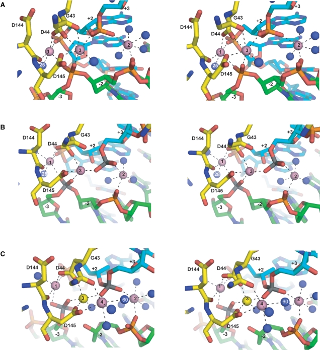
Figure 2.
Stereo views of metal coordinations at the I-SceI/DNA active sites. (A) I-SceI uncleaved DNA complex. (B) I-SceI nicked top strand complex. (C) I-SceI nicked bottom strand complex. Ca2+ are depicted as pink spheres and the Na+ as a yellow sphere. Ordered water molecules are depicted as blue spheres. The scissile phosphoruses are colored gray. Metal coordination spheres are indicated by black dotted lines. The protein backbone is colored yellow, and the top and bottom DNA backbones are colored cyan and green, respectively.

