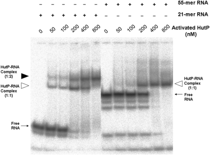
Figure 3.
Gel-shift assay showing HutP–RNA complexes. All reactions were carried out in binding buffer (15 mM HEPES pH 7.5, 30 mM NaCl) with 20 nM of RNA (labeled RNA 104 c.p.m.) containing 10 mM of l-his and Mg2+ ion. To this reaction mixture, various amount of purified HutP protein (to a final concentration of 50–800 nM) were added, and after an incubated for 15 min, the reactions were fractionated by 8% native PAGE. The positions of the free and complex RNA (protein to RNA, 1:1 ratio) are indicated by an arrow and an open arrowhead, respectively. The 21-mer RNA complex (protein to RNA, 1:2 ratio) is indicated by a filled arrowhead.

