
Figure 1. PMN pericellular proteolysis.
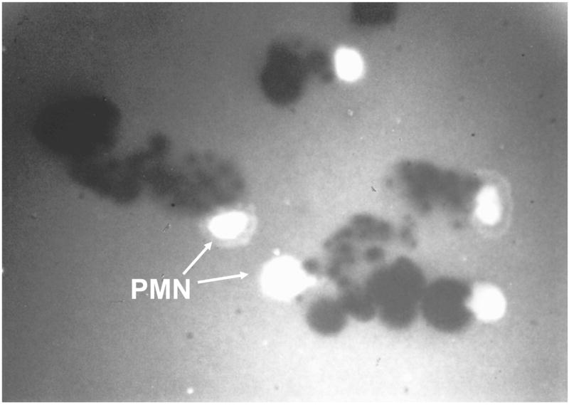
PMNs were incubated for 45 min. at 37°C on FITC-conjugated fibronectin which had been coated on tissue culture plates, and then opsonized. PMNs were bathed in 100% autologous serum which contains micromolar concentrations of TIMPs and serine proteinase inhibitors. Note that PMNs degrade fibronectin substrate as they migrate over it. However, fibronectin degradation is localized to the pericellular environment of the migrating PMNs by the inhibitors present in the bathing medium (arrows). Thus, physiologic proteinase inhibitors present in serum cannot block PMN pericellular proteolytic activity. When cells are bathed in inhibitor free buffers, the FITC-conjugated FN is completely degraded (not shown). One mechanism leading to this inhibitor-resistant pericellular proteolysis is expression of proteinases on the PMN surface in inhibitor-resistant forms which has been demonstrated for several serine and metallo-proteinases families.