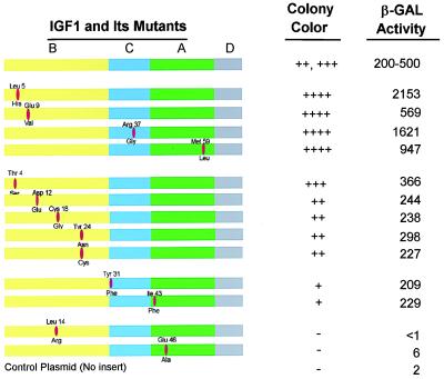
Figure 4.
Quantitation of the interaction of IGF-1 mutants with cysteine-rich domain of IGF-1 receptor. The structure of the IGF-1 is presented schematically with the B, C, A, and D domains. The solid bars indicate the position of the mutation and the number is the amino acid number. The amino acid above the bar is the native sequence; that below the bar is the mutation. Colony color and β-galactosidase activity were measured as described in the legend of Fig. 1.

