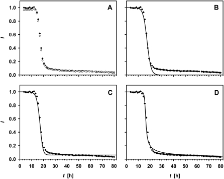
FIGURE 3.
(A) 1H liquid-state NMR signal intensity of the His-1 ɛ1 (open diamonds) and Ala-19 β (solid diamonds) protons as a function of time after dissolving the sample, and the attempts to fit a logistic function to the Ala-19 Hβ data without (B, line) (Eq. 4) and with (C, line) (Eq. 5) a backward rate. (D) This shows the fitting to the data of a model (line) (Eq. 9), where the trimer is the only fibril-forming species.

