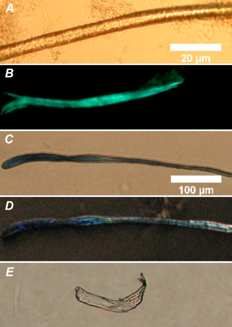
FIGURE 6.
Fibers formed by reacting DPPC with phospholipases A2. Brightfield microscopy image (A), and fluorescence image after ThT staining (B) for bee venom PLA2, DIC microscopy images for porcine pancreatic PLA2 (C) and porcine pancreatic PLA2 zymogen, proPLA2 (D), and birefringence for porcine pancreatic PLA2 after Congo red staining (E, the same fiber as in C). Lipid and the indicated enzymes were mixed at ambient temperature (∼24°C) in 5 mM HEPES, and 0.1 mM EDTA pH 7.4, to yield final concentrations of 100 μM and 0.2 mg/mL, respectively. The scale bars correspond to 20 μm (A and B) and 100 μm (C–E).

