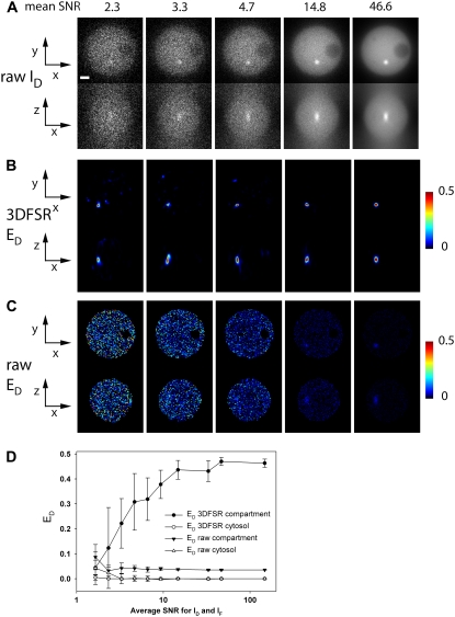
FIGURE 3.
3DFSR improves accuracy and sensitivity over conventional FRET microscopy at all noise levels. Data simulated with various noise levels were reconstructed with 15 iterations of 3DFSR. 3DFSR provided accurate reconstruction of the FRET-positive compartment as seen by the ED signals approaching 0.50 (A), whereas direct application of FRET stoichiometry only allowed detection of the FRET-positive compartment at high SNR (SNR ∼14.8). (D) The mean and standard deviations from 10 realizations for the FRET-positive compartment and a region in the cytosol indicated that 3DFSR could accurately recover the FRET signal in the compartment (solid circles) while correctly suppressing noise to zero (open circles), whereas conventional FRET stoichiometry measured the FRET-positive compartment to be well below the expected 0.50 at all SNR levels (solid triangles) and gave cytosol signals near zero (open triangles). Diminishing values of ED with decreasing SNR correlated with smaller size of the reconstructed FRET-positive compartment. Scale bar is 1 μm. xy and xz slices are from the same planes as in Fig. 1.

