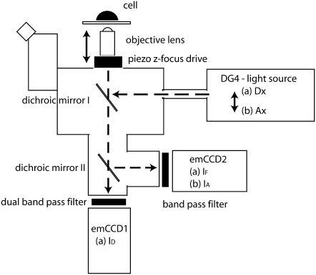
FIGURE 4.
Diagram of the 4D-FRET microscope. The 4D-FRET microscope was an inverted microscope optimized for rapid acquisition of three or four fluorescence images at multiple planes of focus. Broken lines indicate light paths. A water-immersion objective lens with correction collar minimizes spherical aberration in live samples. The z focus is controlled by a piezo drive that moves the objective lens. A fast light source switched rapidly between two illuminations: (a) donor excitation (Dx) and (b) acceptor excitation (Ax). The switching of wavelengths and the z axis movement of the piezo device are the only moving parts and both require 1–10 ms to change. Excitation light is reflected onto the sample by dichromatic beamsplitter I. Sample fluorescence passes through beamsplitter I to a second dichromatic beamsplitter contained within the microscope via custom-built optics and optomechanics. Dichromatic beamsplitter II transmits CFP emission to emCCD1 while reflecting the YFP emission to emCCD2. When illumination a is in place, emCCD1 records the ID image and emCCD2 simultaneously records the IF image. When illumination b is in place, emCCD2 records IA. Device streaming was used to switch illumination and move the z focus (requiring a total of ∼10 ms) during the read time after each exposure, and the next image is captured. The cameras are frame-transfer and run synchronously in streaming mode.

