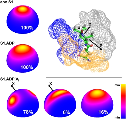
FIGURE 5.
Orientational distribution of the spin label within myosin in the apo, ADP, and ADP Vi biochemical states, based on the model of slow restricted motion. The distribution of z axis of the spin label is color-coded. X axis in spin-label distribution of the S1.ADP.Vi state (M** structural state) is marked to indicate the symmetry about x axis. (Inset) Position of a spin label within apo S1, after Monte Carlo minimization, showing converter domain (blue), N-terminal domain (gray), and relay helix (yellow). The axes of spin label molecular frame are x, y, and z. The colored mesh indicates myosin atoms located at a distance <0.5 nm relative to the spin label.

