
Figure 9.
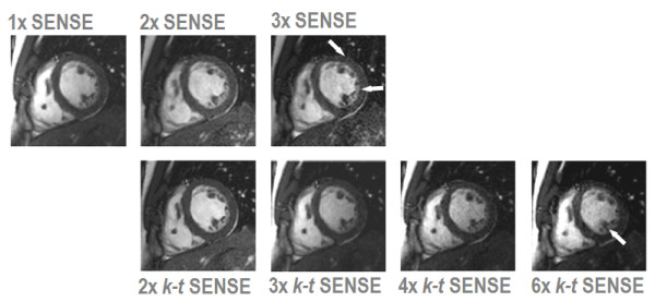
Comparison of clinical 2D cine imaging in the short axis orientation acquired with SENSE and k-t SENSE using a 6-element coil array. Each image depicts the end-diastolic frame of separate breath-hold cines acquired with 1× to 3× SENSE and 2× to 6× k-t SENSE. 2× SENSE provides excellent image quality. With 3× SENSE, the SNR reduction and image artifacts become apparent (arrows). k-t SENSE up to 4-fold net acceleration results in good image quality. At 6× k-t SENSE, slight blurring of the endocardial wall and papillary muscles is visible (arrow).