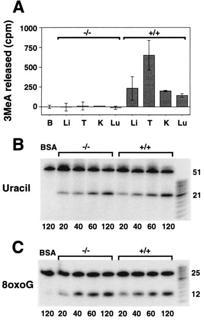
Figure 2.
3MeA, uracil, and 8-oxoG DNA glycosylase activity in mouse tissue extracts. (A) 1,000 μg (BSA, B; liver, Li; and kidney, K), 500 μg (lung, Lu) or 250 μg (testes, T) of protein extracts from Aag +/+ or Aag −/− tissues were incubated in triplicate reactions for 5 hr at 37°C with [3H]methyl-N-nitrosourea-treated calf thymus DNA. 3MeA was separated from other bases by descending paper chromatography. The cpm associated with 3MeA per mg protein extracts are indicated for the average of two or three independent mice. Error bars represent standard deviations. (B) Time-dependent release of site-specific uracil from 5′ 32P-labeled double-stranded oligonucleotides by Aag −/− and Aag +/+ liver extracts. BSA (100 μg) was incubated with the oligo substrate (first lane). Remaining lanes each contain 100 μg of either Aag −/− or Aag +/+ liver extract. After incubation at 37°C for the times indicated in min beneath each lane, oligonucleotides were chemically cleaved at abasic sites and analyzed by denaturing PAGE. DNA glycosylase activity is indicated by the appearance of a 21-mer. (C) Time-dependent release of site-specific 8oxoG from 5′ 32P-labeled double-stranded oligonucleotides by Aag −/− and Aag +/+ testes extracts. BSA (75 μg) were incubated with the 8oxoG containing oligonucleotides (first lane). Remaining lanes each contain 75 μg of either Aag −/− or Aag +/+ testes extracts. Samples were analyzed as described in B. DNA glycosylase activity is indicated by the appearance of a 12-mer.

