
Figure 2. Murine Adhfe1 is a 50 kDa Protein with Mitochondrial Localization.
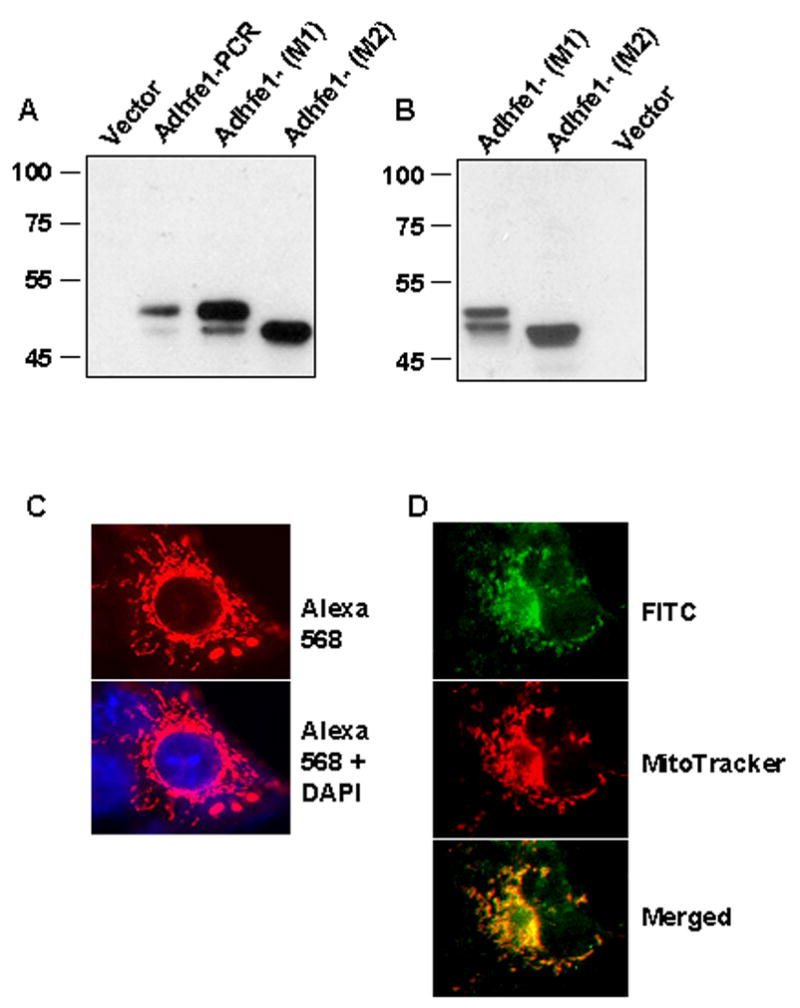
A: In vitro transcription and translation of Adhfe1. Adhfe1 PCR product and Adhfe1 expression constructs with sequence starting at the first ATG (M1) or lacking the first ATG and starting at the second ATG (M2) were transcribed and translated as described under “Materials and Methods” with empty vector used as a negative control. The in vitro translation products were analyzed by Western blot using anti-HA primary antibody. Asterisk indicates major Adhfe1 protein species and the arrow indicates minor Ahdfe1 protein species that is attributed to translation from second ATG when the M1 construct was used as template. B: Expression of Adhfe1 protein. COS cells were transfected with empty pcDNA3.1 vector or a HA tagged Adhfe1 constructs and analyzed by Western blot using anti-HA primary antibody. C: Localization of Adhfe1 protein. COS cells were transfected with HA-tagged Adhfe1construct. Immunostaining was carried out with anti-HA primary antibody and AlexaFluor 586-conjugated secondary antibody, resulting in a red signal (top panel). The lower panel shows merged image from AlexaFluor 586 and DAPI nuclear staining. D. Adhfe1 Localizes to Mitochondria. COS cells were transfected with an HA-tagged Adhfe1construct. Immunostaining was carried out with anti-HA primary antibody and FITC-conjugated secondary antibody resulting in a green signal for Adhfe1 (top panel). Middle panel shows MitoTracker CMX Ros staining, visualized as red signal and the lower panel the merged image where yellow signal indicates overlap of Adhfe1 and MitoTracker staining. Protein expression studies are representative of n=2.