
Figure 3. Upregulation of Adhfe1 is Closely Tied to White and Brown Adipogenesis.
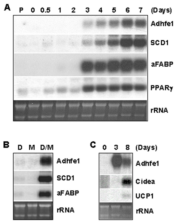
A: Adhfe1 upregulation during 3T3-L1 differentiation. 5 μg of total RNA from 3T3-L1 preadipocytes (P), confluent 3T3-L1 cells before induction of adipogenesis (day 0) and indicated time points after induction of adipogenesis were analyzed by Northern blot using Adhfe1, SCD1, aFABP, and PPARγ cDNA probes. B: Regulation of Adhfe1 transcript expression. Post-confluent 3T3-L1 cells were treated with 1 μM dexamethasone (D), 0.5 mM MIX (M), or 1 μM dexamethasone and 0.5 mM MIX (D/M) for 48 h and harvested at day 5. Northern blot analysis was performed with murine Adhfe1, SCD1, and aFABP probes. C: Adhfe1 transcript expression in brown adipogenesis. Brown preadipocytes were differentiated as described in “Materials and Methods” at confluence (day 0). Brown adipocytes were harvested at 3 and 8 days and Northern blot analysis was performed with murine Adhfe1, Cidea, and UCP1 probes. For A-C, the EtBr staining of rRNA is shown as a gel loading control. Northern blots are representative of n=2.