
Figure 3.
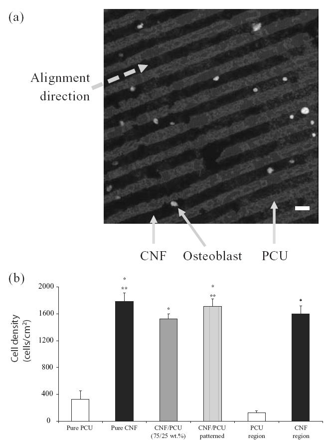
(a) Optical image of selective osteoblast adhesion on carbon nanofiber (CNF) patterns on polycarbonate urethane (PCU) after 2 days of culture (bar = 20 μm). (b) Increased osteoblast adhesion on patterned CNF–PCU compared with nonpatterned CNF–PCU. Values are mean +/– standard error of mean; n = 7; *p < 0.01 (compared with pure PCU or PCU regions) and **p < 0.01 (compared with PCU–CNF not patterned).