
Figure 6.
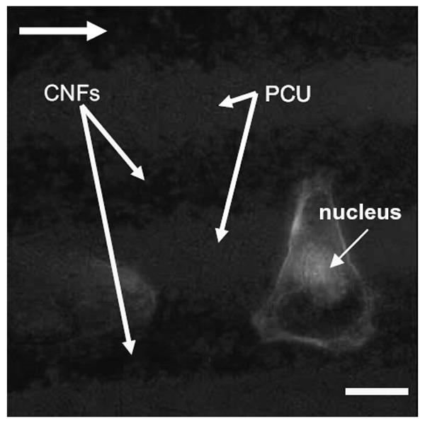
Fluorescence microscopy image of an osteoblast adherent at the edge of a carbon nanofiber (CNF) pattern in polycarbonate urethane (PCU). Note that for one osteoblast, f-actin filaments stretch across the PCU pattern to an adjacent CNF pattern. Bar = 10 μm; culture time = 2 days; arrow shows direction of CNF patterns.