
Figure 2.
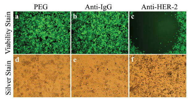
Antibody conjugated nanoshells bound to cells when the appropriate antigen was present. SK-BR-3 cells were incubated with PEG-coated nanoshells (a, d), anti-IgG conjugated nanoshells (b, e), and anti-HER2 conjugated nanoshells (c, f). Following laser exposure, a region of cell death corresponding to the laser spot resulted in groups incubated with anti-HER2 conjugated nanoshells (c). Cells incubated with PEGylated or anti-IgG conjugated nanoshells continued to live. Silver staining (d–f) showed maximal binding of anti-HER2 conjugated nanoshells to the SK-BR-3 cells (f). Laser spot is 1.5 mm wide.
Abbreviations: HER2, epidermal growth factor receptor; PEG, polyethylene glycol.