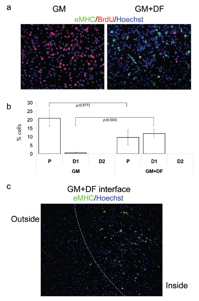
Figure 1.

Geometric control of terminal myogenic differentiation in GM. Myogenic progenitor cells have been plated at 50% confluency in chamber slides in GM.
(a) GM with unmodified ECM (GM) and GM with DF-modified ECM (GM + DF). (b) Quantification of P, differentiated cells with less than 2 nuclei (D1 = early stage of differentiation), and differentiated cells with more than 2 nuclei (D2 = later stage of differentiation). On unmodified ECM, there is a significantly higher percentage of proliferative cells vs differentiated cells. Looking at cells grown on DF-modified ECM, we see higher numbers of differentiated cells, but most cells do not form multinucleated myotubes. (c) The boundary between unmodified ECM substrate (outside) and DF-modified ECM (inside). Geometric boundary between DF-modified and unmodified ECM substrate was created as described in Methods. Cells were uniformly seeded throughout the ECM area, cultured for 48 hours and fixed. Immunofluorescence was performed with the indicated antibodies: α-BrdU (red), α-eMHC (green), and Hoechst (blue) was used to label all nuclei. Proliferation (incorporation of BrdU) is observed in GM with unmodified ECM and inhibited proliferation and differentiation (expression of eMHC) is observed in GM with DF-modified ECM. Similar results have been obtained in at least 3 independent experiments. Magnification: (a) 20x; (c) 10x.
Abbreviations for Figures 1-3, Scheme 1, SOM 1: D, differentiated cells; DF, differentiation factors; DM, differentiation-promoting medium; ECM, extracellular matrix; GM, growth-promoting medium; GF, growth factors; NM, neutral medium; P, proliferating cells; PBS, phosphate buffered saline.
