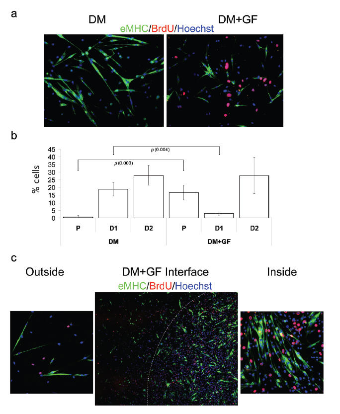
Figure 2.

Geometric delay of myotube formation in DM by locally embedded growth factors. Myogenic progenitor cells have been plated at 50% confluency in chamber slides in DM. Cells were cultured for 48 hours and fixed. Immunofluorescence was performed with the indicated antibodies: α-BrdU (red), α-eMHC (green), and Hoechst (blue) was used to label all nuclei.
(a) DM with unmodified ECM (DM) and DM with GF-modified ECM (DM+GF). There is a clear difference in the fate of cells cultured in DM on unmodified ECM (terminally differentiated, multinucleated myotubes) vs on GF-modified ECM (higher numbers of proliferative cells and smaller myotubes). (b) Quantification of P, early stage-differentiated cells with less than 2 nuclei (D1) and later stage-differentiated cells with more than 2 nuclei (D2). Cells cultured on unmodified ECM in DM show low percentage of proliferating cells and high percentage of differentiated cells. Alternately, when cultured on GF-modified ECM, cells show higher percentage of proliferating cells and lower numbers of differentiated cells. (c) The boundary between unmodified ECM substrate and DF-modified ECM is shown (DM + GF interface). Magnified photographs (20x) of cells seeded on unmodified ECM (outside) and on GF-modified ECM (inside) areas of the culture plate are also shown. Cells were originally seeded at uniform confluency; however, as expected, cells adherent to the GF-modified ECM proliferated at a higher rate, resulting in a higher number of cells compared with those adherent to control ECM. Similar results have been obtained in at least 3 independent experiments. Magnification: (a) 20x; (c) “Outside”, “Inside” 20x; (c) “Interface” 10x.
