Figure 3.

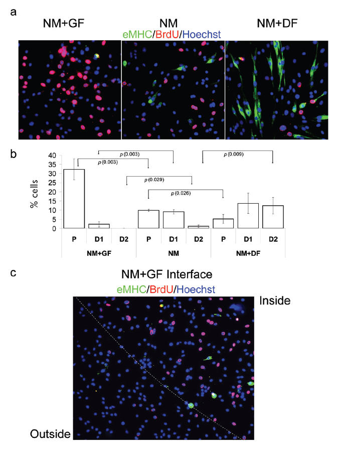
Geometric control of proliferation or terminal differentiation in NM. Myogenic progenitor cells have been plated at 50% confluence in chamber slides in NM for 48 hours. Immunofluorescence was performed with the indicated antibodies: α-BrdU (red), α-eMHC (green), and Hoechst (blue) was used to label all nuclei.
(a) Cells cultured in NM on unmodified ECM substrate (NM) show no distinct tendency towards proliferation or differentiation. When cultured on GF-modified ECM (NM + GF) cells proliferate (BrdU incorporation) without any tendency to form myotubes and differentiate, ie, eMHC+. Conversely, when exposed to DF-modified ECM (NM + DF), cells terminally differentiate (eMHC+) and form myotubes while proliferation is reduced. (b) Quantification of P, early stage-differentiated cells with less than 2 nuclei (D1), and later stage-differentiated cells with more than 2 nuclei (D2). When cultured in NM on unmodified ECM, cells infrequently differentiate and have slow proliferation rate. However, when cells are plated on GF-modified ECM (NM + GF) there is a much larger percentage of proliferating cells; and when they are plated on DF-modified ECM (NM + DF) there are higher percentages of not only eMHC+ differentiated cells but also yield higher percentages of multinucleated myotubes (D2). (c) Boundary between GF-modified ECM and unmodified ECM. There are higher numbers of proliferating cells on GF-modified ECM (inside) than on the unmodified ECM (outside). Similar results have been obtained in at least 3 independent experiments. Magnification: (a) 20x; (c) 10x.
