SOM 1.

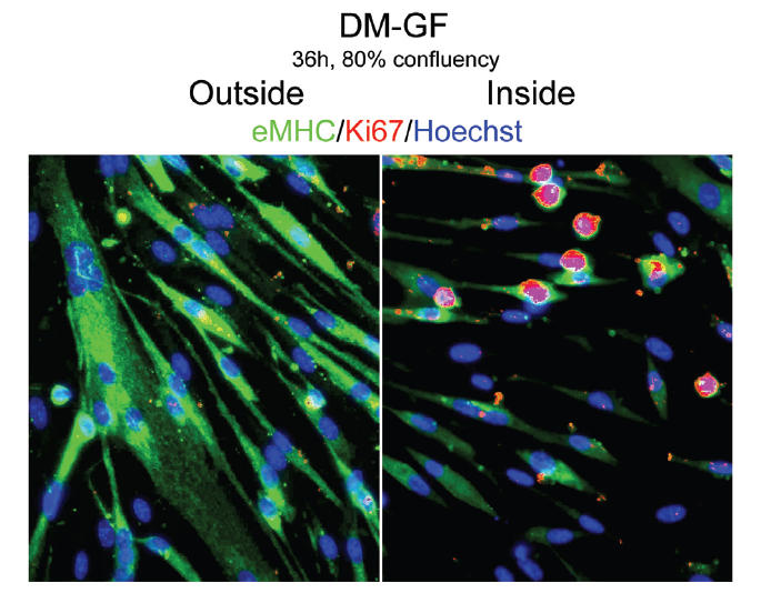
Delay of myogenic differentiation by locally embedded growth factors into ECM under high cell density (80% confluency). Myogenic progenitor cells have been plated at 80% confluency in chamber slides in DM for 36 hours. Immunofluorescence was performed after fixation with the indicated antibodies: α-Ki67 (red), α-eMHC (green), and Hoechst (blue) was used to label all nuclei. As specified in Table 1, GF were embedded into ECM in geometric fashion, as shown in Scheme 1. Consistent with the control DM shown in Figure 2a-DM, cells outside the geometric boundary (outside) form eMHC-positive robust myotubes and do not proliferate. Myogenic cell differentiation is diminished, as indicated by the lower number of nuclei per myotubes, and some Ki67+ proliferating cells persist inside the geometric boundary (inside). Thus, GF embedded in the ECM are capable of diminishing differentiation even when cell numbers increase, but differentiation seems inescapable despite initial placement of GF into ECM. Magnification: 10x.
