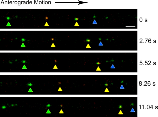
Figure 6. Imaging transport of light and heavy particles inside the microfluidic chamber.
Cell bodies in the somal compartment were infected with PRV 181, a recombinant expressing an mRFP-VP26 (capsid) fusion and a GFP-VP22 (outer tegument) fusion. Time-lapse images were taken of axons within the microgrooves at 11–14 h postinfection using the Leica SP5 confocal microscope. Green puncta of light particles containing GFP-VP22 without mRFP-VP26 (green and blue arrows) and yellow punta of heavy particles containing both GFP-VP22 and mRFP-VP26 (yellow and orange arrows) were seen moving in the anterograde direction. Both light and heavy particles displayed heterogeneity in GFP fluorescence. For a complete time-lapse recording, see Movie S2 in the supporting information. Bar: 2 μm.

