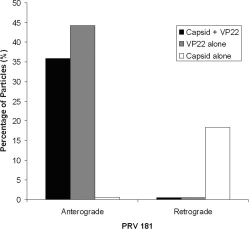
Figure 7. Transport direction of capsids and VP22 in axons during viral egress.
Cell bodies in the somal compartment were infected with PRV 181, a recombinant expressing an mRFP-capsid fusion and a GFP-VP22 fusion. Viral particles traveling in axons within the microgrooves were tracked from 11–14 h postinfection. The fraction of capsids transported with (black) or without (white) VP22 and the fraction of VP22 moving in the absence of capsids (grey) are shown as percentages of the total particles tracked.

