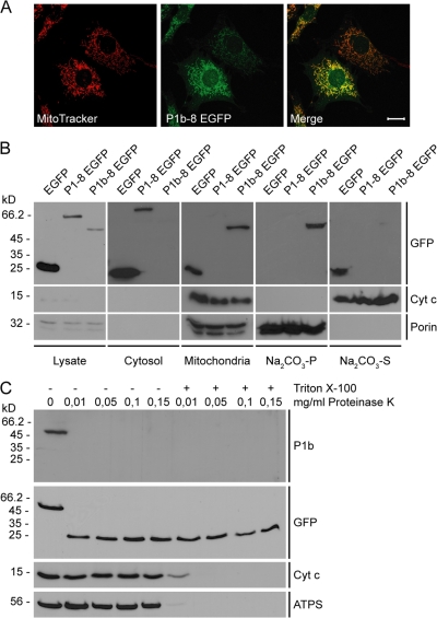
Figure 1.
Submitochondrial localization of P1b. (A) Colocalization of P1b–8 EGFP with mitochondria in stably transfected P0 fibroblasts visualized using anti-GFP antibodies and MitoTracker. Bar, 20 μm. (B) Cell lysates and cytosolic and mitochondrial fractions were obtained from fibroblasts stably expressing P1b–8 EGFP, P1–8 EGFP, or EGFP. Mitochondria were extracted with sodium carbonate, and insoluble pellet (Na2CO3-P) and soluble supernatant (Na2CO3-S) fractions were subjected to immunoblotting. White lines indicate that intervening lanes have been spliced out. (C) Mitochondria were left untreated or lysed with Triton X-100 before the addition of indicated amounts of proteinase K.

