
Figure 2.
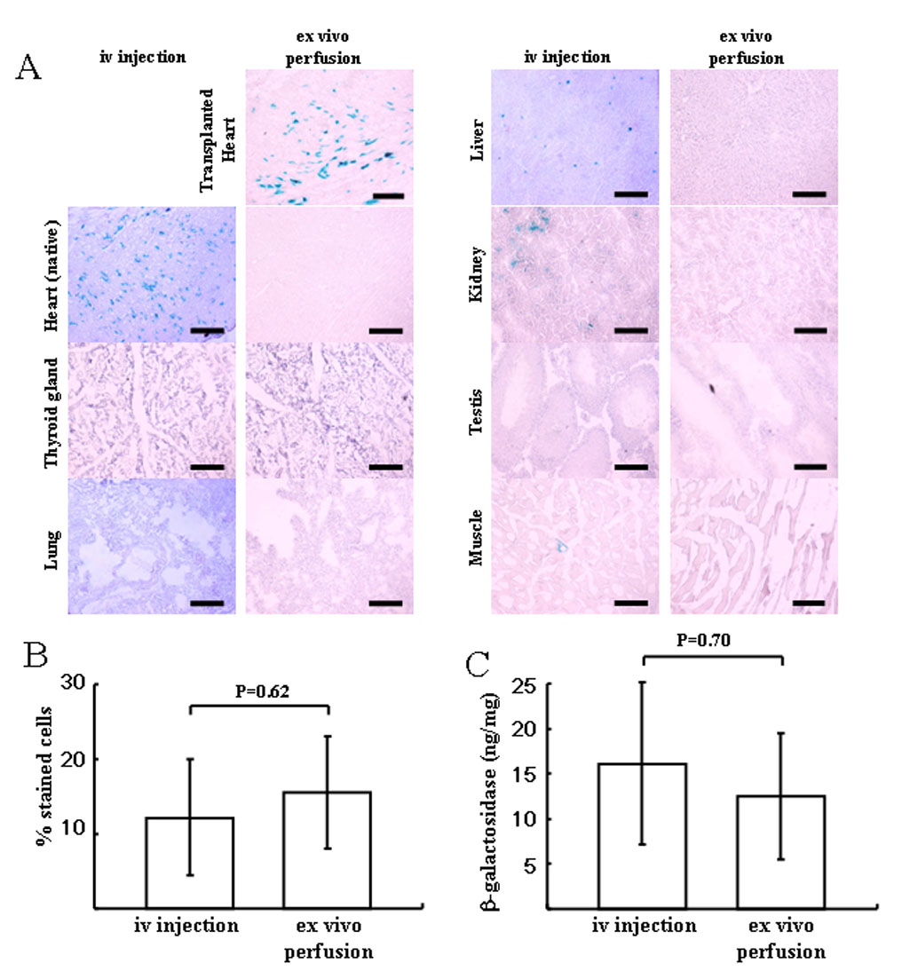
Tissue distribution of β-galactosidase-positive cells in the iv injection group and ex vivo perfusion group using AAV9-CMV-lacZ (1×10¹² vg). (A) Representative microphotograph in each group. Each section was stained with X-gal and light hematoxylin. No positive cells were observed in thyroid gland, native heart, liver, kidney, skeletal muscle (quadriceps femoris) or testis of the recipient in ex vivo perfusion group. In the iv injection group, β-galactosidase positive cells were in heart>liver>kidney>muscle. Scale bar represents 100um. (B) Comparison of efficacy of lacZ gene transduction between the native heart in iv injection group and the transplanted heart in ex vivo perfusion group. (C) Total β-galactosidase antigen level. No significant difference was seen between two groups in the transduction efficiency. Values are means ± standard deviation.