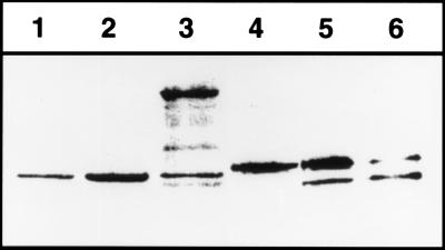
Figure 2.
Proteolysis of SecA410 in vivo. To monitor cleavage of SecA410 by TEV protease, a Western blot was prepared by using cells of ME7000 expressing either wild-type SecA (lane 1), wild-type SecA expressed from pMF8 (lane 2), secATnTIN410 (lane 3), SecA410 resulting from a cutback of TnTIN with NotI (lane 4), or SecA410 plus TEV protease expressed from pMM13 (lanes 5 and 6) after overnight growth in Luria–Bertani. The growth medium was without (lane 1–5) or supplemented with 100 μM isopropyl β-d-thiogalactoside (lane 6) to induce TEV protease. To detect SecA, polyclonal antibodies against SecA were used.

