
Figure 4.
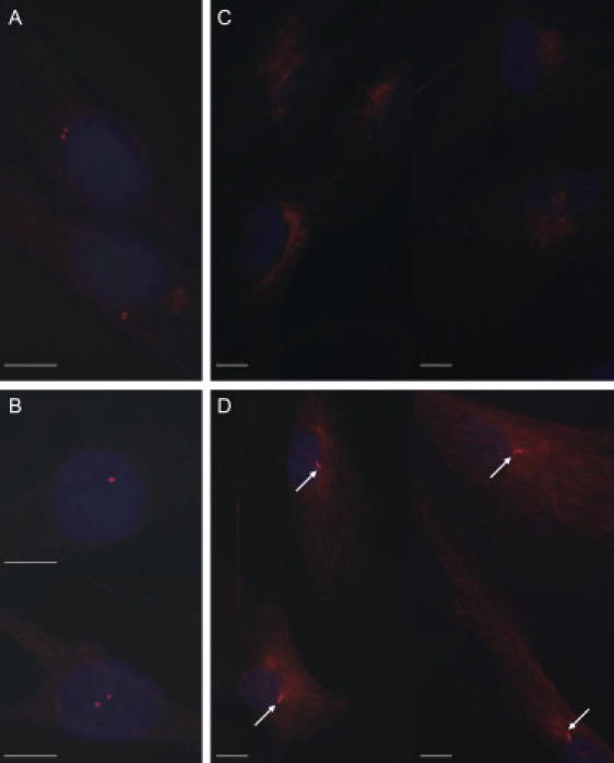
Immunofluorescence Staining of Centrioles and Cilia
Centrioles can be visualized with γ-tubulin staining in fibroblast cells of a patient (A) and a healthy control (B) (magnification 100×). Staining with acetylated tubulin shows that cilia are missing in the patient cells (C) but can be observed in control cells (D) (white arrows, magnification 63×). Scale bar represents 10 μm.