
Figure 1.
Genetic Analysis of the Mutation in NDUFA2
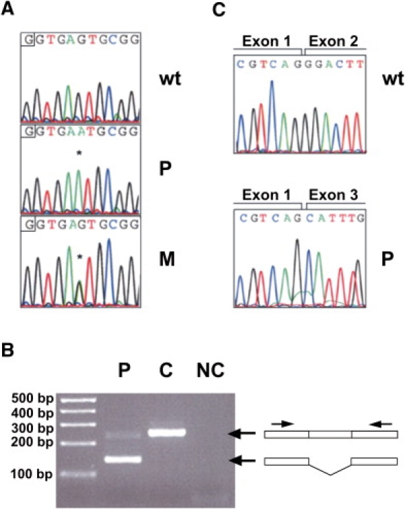
(A) Electropherograms showing the normal sequence of NDUFA2 (top) and the nucleotide change in fibroblasts of the patient (middle) and the mother of the patient (bottom). Asterisks represent the nucleotide substitution.
(B) PCR analysis of the mRNA showed a product of approximately 250 base pairs with control RNA, whereas PCR analysis with patient RNA resulted in a fragment of about 150 base pairs. Notably, a small amount of wild-type product was still present.
(C) Sequence analysis of the PCR fragments of (B) showed a normal exon boundary between exons 1 and 2 in the control (top) and skipping of exon 2 in the patient (bottom).
Abbreviations are as follows: wt = wild-type, p = index patient, M = mother of patient, C = control, and NC = negative control.