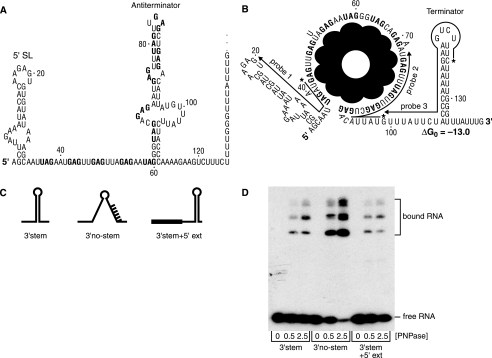
FIGURE 1.
Sequence and secondary structure of trp leader RNA without (A) and with (B) bound TRAP. Numbering is from the natural start site of trp transcription. GAG and UAG triplet repeats at which TRAP binds are in bold. 5′-SL denotes the 5′-stem-loop structure. In B, lines indicate complementarity of probes 1, 2, and 3 to trp leader RNA sequences, with the 5′-labeled end of the probe indicated by an asterisk. The ΔG0 (kcal mol–1) for the trp leader RNA 3′-end structure, calculated according to the default conditions of the mfold version 3.2 site, is shown. C, schematic diagrams of RNA oligonucleotides used to probe RNase J1 activity on the 3′-terminal fragment. D, binding of purified PNPase to RNA oligonucleotides. Concentration (nm) of PNPase is indicated below each lane.

