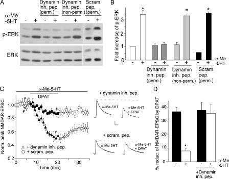
FIGURE 7.
The counteractive effect of 5-HT2A/C on 5-HT1A regulation of NMDAR function involves clathrin/dynamin-mediated endocytosis. A, Western blots of phospho-ERK and total ERK in PFC cultures treated with or without α-Me-5HT (20 μm, 3 min) in the absence or presence of the cell-permeable (myristoylated) or non-permeable dynamin inhibitory peptide (both 50 μm), or a cell-permeable scrambled control peptide (50 μm). This scrambled peptide was fused with the protein transduction domain of the human immunodeficiency virus TAT protein (YGRKKRRQRRR, 53), which rendered it cell-permeable. All peptides were added 30 min before α-Me-5HT treatment. B, quantification of phospho-ERK under different conditions. Each point represents mean ± S.E. of 4–5 independent experiments. *, p < 0.001, ANOVA. C, plot of normalized peak NMDAR-EPSC showing the effect of 8-OH-DPAT (20 μm) in the presence of α-Me-5HT (20 μm) in cells dialyzed with the dynamin inhibitory peptide (50 μm) or a scrambled control peptide (50 μm). Inset, representative traces of NMDAR-EPSC taken at time points denoted by #. Scale bars: 100 pA, 10 ms. D, cumulative data (mean ± S.E.) summarizing the percentage reduction of NMDAR-EPSC by 8-OH-DPAT in the absence or presence of α-Me-5HT in neurons injected with or without the dynamin inhibitory peptide. *, p < 0.001, ANOVA.

