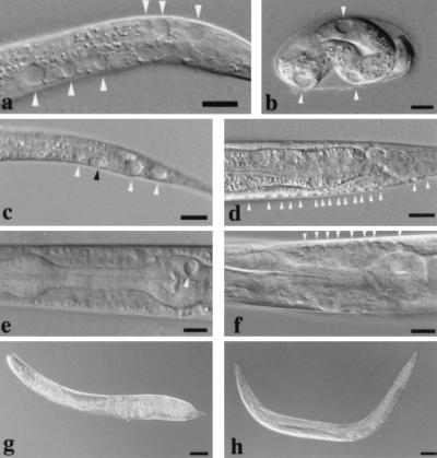
Figure 1.
Effects of ectopic expression of mec-4(d). Animals are oriented with anterior to the left and dorsal to the top, except for embryos. (a–f) Nomarski differential interference contrast microscopy images (Bar = 10 μm). (g–h) Bright-field images (Bar = 50 μm). Most prominent vacuoles, which typically appear as crater-like, swollen, membrane-bound units, are highlighted by arrows. Vacuoles may be evident in several different focal planes, and thus not all appear in sharp focus. In early stage degenerations, swollen nuclei can be observed within cells (for example, c, black arrow; see ref. 8). (a) Extensive vacuolation in a heat shocked L1 animal harboring phsp-16mec-4(d). (b) Heat shocked post-pretzel stage embryo harboring phsp-16mec-4(d). (c) Ventral cord neurons swelling in the posterior of an L1 stage animal harboring punc-4mec-4(d); black arrow indicates a cell with a clearly swollen nucleus inside. (d) Small vacuolated regions in the hypodermis of an L2 animal harboring pmec-5mec-4(d). Note that hypodermal vacuoles are always small and nuclear inclusions within these vacuoles have not been observed. (e) Vacuolation of a pharyngeal muscle in an L2 stage animal harboring pmyo-2mec-4(d). (f) punc-54mec-4(d)-induced swelling of body wall muscle cells near the pharynx in an L3 stage animal. Indicated are several vacuoles along the dorsal side; the ventral side also exhibited small vacuoles in this animal that are not fully in focus. In body wall muscle, individual cells often appear to have multiple small vacuoles as shown. (g) The hypercontracted phenotype of an animal bearing the punc-54mec-4(d) transgene. (h) A mec-6(u450) mutant bearing the punc-54mec-4(d) transgene is not dramatically hypercontracted.

