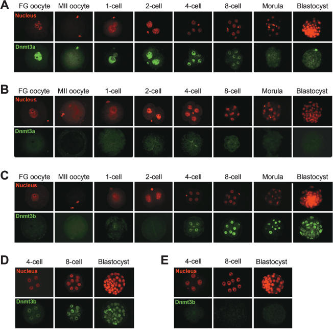
Figure 1.
Expression and subcellular localization of Dnmt3a and Dnmt3b in mouse oocytes and preimplantation embryos. (A) Immunostaining of wild-type FG oocytes, MII oocytes, and preimplantation embryos with an anti-Dnmt3a antibody. Dnmt3a signals (green) were detectable in the nucleus of FG oocytes and embryos from the one-cell through to the eight-cell stage. Dnmt3a was diffusely present in the cytoplasm of MII oocytes. Small intense signals represent the nuclei of pole bodies. (B) Absence of detectable Dnmt3a in oocytes and preimplantation embryos from [Dnmt3a2lox/2lox, Zp3-Cre] females. This confirms the maternal origin of the protein detected in the wild-type embryos. (C) Immunostaining of wild-type oocytes and preimplantation embryos with an anti-Dnmt3b antibody. Dnmt3b (green) was not detectable in oocytes, one-cell embryos, or two-cell embryos and became detectable in the later stages. (D) Zygotic production of Dnmt3b in preimplantation embryos. Dnmt3b was detected in embryos obtained from [Dnmt3b2lox/2lox, Zp3-Cre] females crossed with wild-type males. (E) Absence of detectable Dnmt3b signals in embryos obtained from [Dnmt3b2lox/2lox, Zp3-Cre] females crossed with [Dnmt3b2lox/1lox, Tnap-Cre] males. The cell nucleus was counterstained with propidium iodide (PI) (red).

