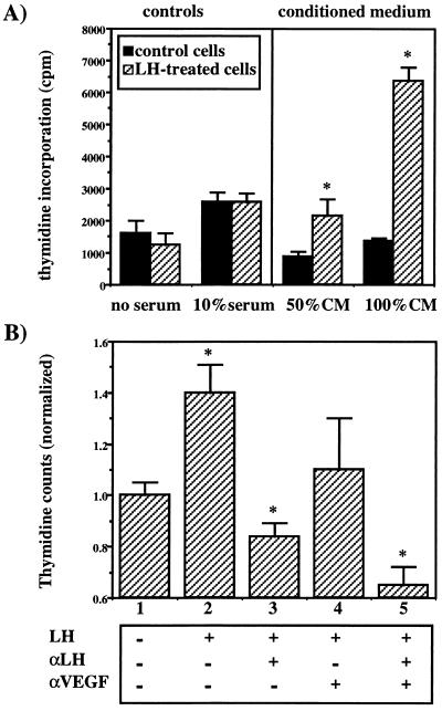
Figure 3.
The effect of medium conditioned by LH-stimulated ovarian cancer cells on endothelial cell proliferation in vitro. (A) Subconfluent MLS cells were cultured in 25-cm2 flasks for 48 h in the medium of the endothelial cells. Half of the flasks were supplemented with hLH (1 μg/ml). The conditioned medium (CM) was collected, filtered at 0.2 μm, and applied immediately on BCE and A21 endothelial cells for a 30-h incubation. Direct stimulating effect of LH on the endothelial cells was ruled out by incubating the cells with or without LH as shown in the left side of A. Cell proliferation was monitored by 3H-thymidine incorporation. Note the dose-dependent enhancement of endothelial cell proliferation in hLH-conditioned medium (P < 0.05). (B) MLS cells were incubated in the presence or absence of hLH and neutralizing antibodies for LH (αLH) for 48 h. The conditioned medium was applied as in A on the endothelial cells in the presence or absence of neutralizing antibodies for VEGF (αVEGF). Cell proliferation was monitored by 3H-thymidine incorporation. A statistically significant difference (one-tailed t test; ∗, P < 0.05) was observed between: columns 1 and 2 (P = 0.006); columns 2 and 3 (P = 0.002); columns 2 and 5 (P = 0.001); columns 3 and 5 (P = 0.024); and columns 4 and 5 (P = 0.023). For columns 2 and 4, P = 0.08.

