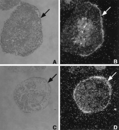
Figure 3.
VE-specific expression of HNF4α and aldoB. Morphological section of HNF4+/+ EBs and in situ hybridization with antisense RNA of HNF4α (B) and aldoB (D) probe. Phase contrast micrograph of 5-μm sections through 14 days ES cell EBs, stained with hematoxylin and eosin (A and C), and the corresponding dark field on the right (B and D). The cuboidal epithelium of the VE is identified by an arrow.

