Figure 1.
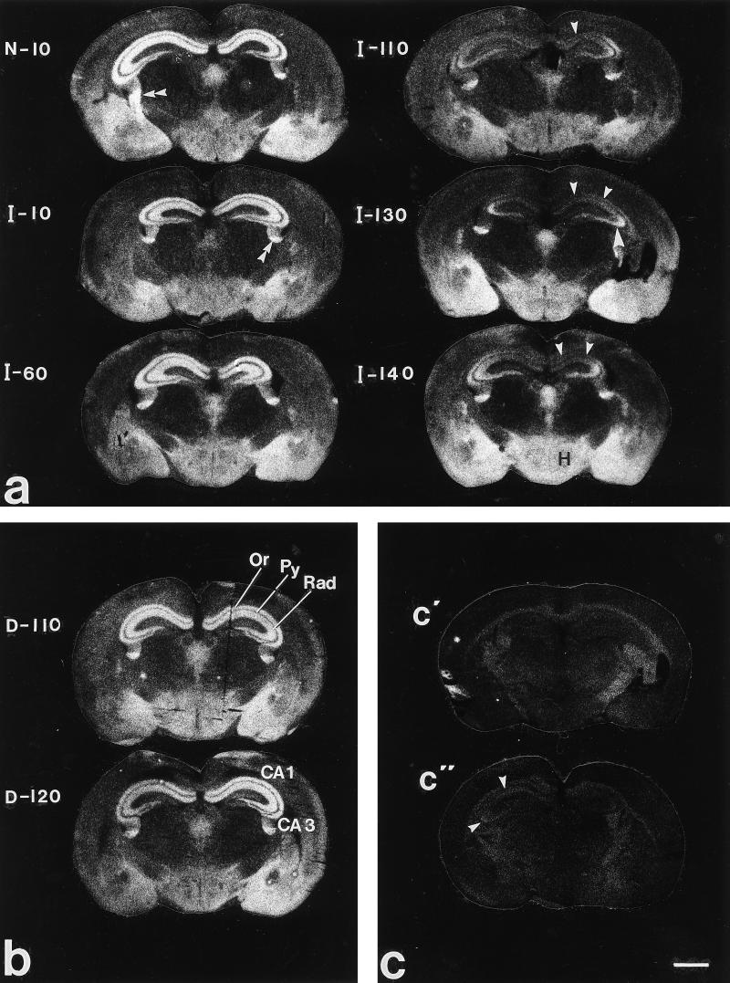
(a–c) X-ray autoradiograms of coronal sections at the level of the dorsal hippocampus of normal (N-10), prion inoculated (I-10 to I-140), and diluent inoculated (D-110, D-120) mouse brains after incubation with 125I-PYY. (c) Coincubation with NPY (c′) or the Y2 agonist NPY (13–36) (c"). (a) No differences can be observed between the control brain and brains 10 or 60 days after prion inoculation. However, 110 to 140 days after inoculation, there is a dramatic decrease in binding both in stratum radiatum and stratum oriens in the CA1 region (arrowheads), as well as in stratum oriens of the CA3 region (big arrow heads). Note lack of apparent differences in the hypothalamus (H). Double arrowheads point to stria terminalis. (b) No changes in binding can be seen in brains injected with diluent. (c) Complete blockage of binding is seen after coincubation with cold NPY (c′) whereas a weak binding can be seen both in the CA1 and CA3 regions (arrowheads) after coincubation with NPY (13–36) (C"). (Bar = 800 μm.) All micrographs have the same magnification.

