Figure 3.—
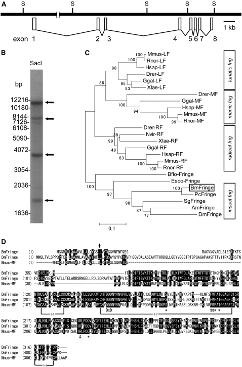
Genomic and phylogenetic characterization of Bombyx fng. (A) The genomic structure of Bombyx fng, which consists of eight exons. S indicates SacI restriction site. (B) Genomic Southern analysis with the fng probe for WT genomic DNA restricted with SacI. The cDNA of fng ORF region is used as the probe (see materials and methods). Arrows indicate four positive bands. The DNA-size markers are shown to the left. (C) Phylogenetic tree of fng based on the amino acid sequences. The numbers at the tree edge represent the bootstrap values. The scale bar indicates the evolutionary distance between the groups. Bombyx fng (BmFringe) is boxed. Respective fng sequences are shown in materials and methods. (D) Amino acids alignment of Fng from Bombyx (BmFringe), Drosophila (DmFringe), and Manic-fng from mouse (Mmus-MF). Black shading with white letters indicates identical amino acid residues. Arrow shows the putative signal peptide cleavage site. DxD show conserved DxD motif. Asterisks and number signs (#) show the putative amino acid residues involved in UDP-GlcNAc binding and fucose-binding, respectively. s-s indicates disulfide bond.

