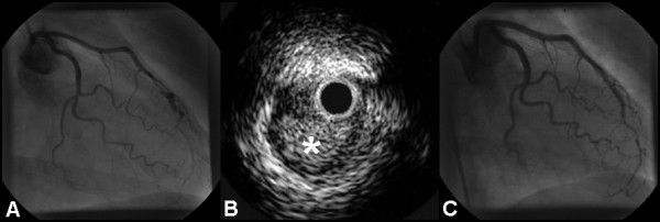
Figure 1.

(A) RAO caudal projection showing dissection of the LAD in the mid-vessel.(B) IVUS examination showing hematoma (asterisk) compressing the true lumen. (C) RAO caudal view of the LAD demonstrating a favourable angiographic appearance following stent deployment.
