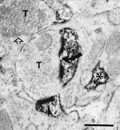
Figure 1.
Electron micrographic localization of rat visual cortex G3PD. Experimental procedures are as stated in the Materials and Methods section. The G3PD antibody was diluted 1:2,000. T marks unlabeled terminals whereas arrowheads mark immunoreactive PSD at a dendritic spine. The large curved arrow points to the perforation along one immunoreactive PSD, whereas the open arrow points to an unlabeled PSD. An asterisk denotes an immunoreactive astrocytic process. (Bar = 500 nm.)

