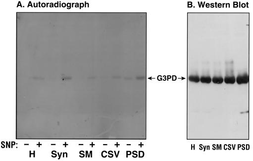
Figure 2.
(A) NO-stimulated [adenylate-32P]NAD incorporation into G3PD in subcellular fractions isolated from adult porcine cerebral cortex. Assays are described in Materials and Methods. PSD (50 μg) and 100 μg each of the other fractions, in 100 μl final volume, including whole homogenate (H), synaptosomes (Syn), synaptic plasma membranes (SPM), and crude synaptic vesicles (CSV), were incubated at 37°C for 15 min. NAD incorporation was performed in the absence (−) or presence (+) of SNP as exogenous source of NO. The mixtures were subjected to SDS/PAGE and then autoradiography. (B) Western blot analysis of the G3PD in the subcellular fractions. To confirm that the radioactive protein in the subcellular fractions was indeed G3PD, Western blot analysis was performed by using specific anti-G3PD antibodies as described.

