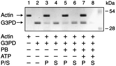
Figure 5.
Effects of autophosphorylation of purified G3PD on subsequent binding to actin. Experiments were performed as described. G3PD was incubated with and without ATP. The mixtures were centrifuged to separate pellets (P) and supernatants (S), subjected to SDS/PAGE and proteins stained with Coomassie brilliant blue. Lanes 1 and 2 are actin and G3PD, respectively. Lanes 3 and 4 are actin and G3PD incubated together. Lanes 5 and 6 are G3PD treated with phosphorylation buffer (PB) without ATP. Lanes 7 and 8 are G3PD treated with the ATP and PB. Molecular weight marker proteins are shown on the right.

