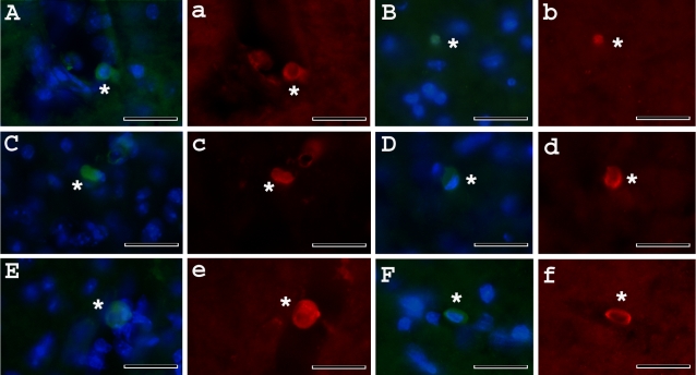
Figure 10. Immunochistochemical staining of MNC hUCB cells in the lumbar spinal cord of G93A mice administered with different cell doses.
MNC hUCB cells were found in the lumbar spinal cord of mice receiving A), B) 10×106; C), D) 25×106; and E), F) 50×106 cells by anti-human nuclei staining (green, asterisks). Merged images are with DAPI. Some a), b), c), d), e), f) MNC hUCB cells expressed Nestin (red, asterisks). Cells in images a, b, c, d, e, f are same in images A, B, C, D, E, F. Scale bar: A–e is 25 µm.

