Fig. 1.
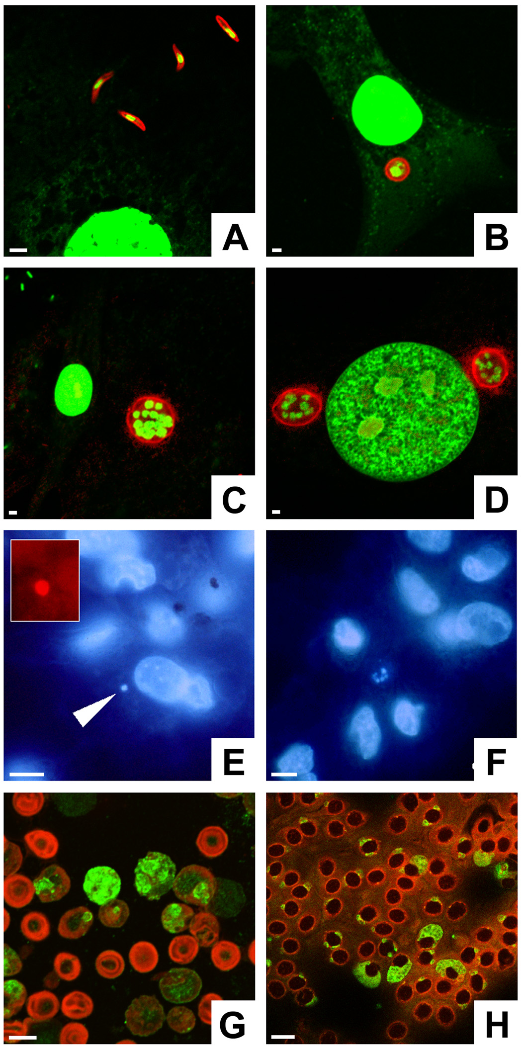
Detection methods for various Plasmodium gallinaceum life cycle stages. A–D) Sporozoites and first generation exoerythrocytic stages can be detected by circumsporozoite protein (CSP) labeling. SL-29 chicken fibroblasts were co-cultivated with sporozoites and fixed after different periods of time. The parasites were labeled with mAb N2H6D5 against P. gallinaceum CSP in combination with goat anti-mouse IgG conjugated to Texas Red (red). The nucleic acid stain YOYO-1 was used to visualize parasite and host cell nuclei (green). Fibroblast cultures contained extracellular sporozoites at 1 h (A), immature trophozoites at 24 h (B), and mature schizonts at 48 h (C). At 48 h, two mature schizonts flank the nucleus of the host cell (D). Note the relatively small diameter of the parasites compared with liver stages from mammalian Plasmodium species, which grow larger than the original host hepatocyte. E, F) Bone marrow macrophage cultures were infected with P. gallinaceum sporozoites and stained with the nucleic acid dye Hoechst 33342. E) An infected cell contains a parasite with a single nucleus (arrow). F) Multi-nucleated schizonts, recognizable by their small nuclei (center), were found after 2 days of co-cultivation. G) Anti-Pb, generated against Plasmodium berghei-infected murine erythrocyte ghosts, reacts with all blood stages of this rodent parasite. Anti-Pb was used in combination with goat anti-rabbit IgG conjugated to fluorescein isothiocyanate (green); the blood cells were counter-stained with Evans blue (red). H) Anti-Pb labels conserved blood stage antigens in P. gallinaceum-infected chicken erythrocytes (green). Bars = 5 µm.

