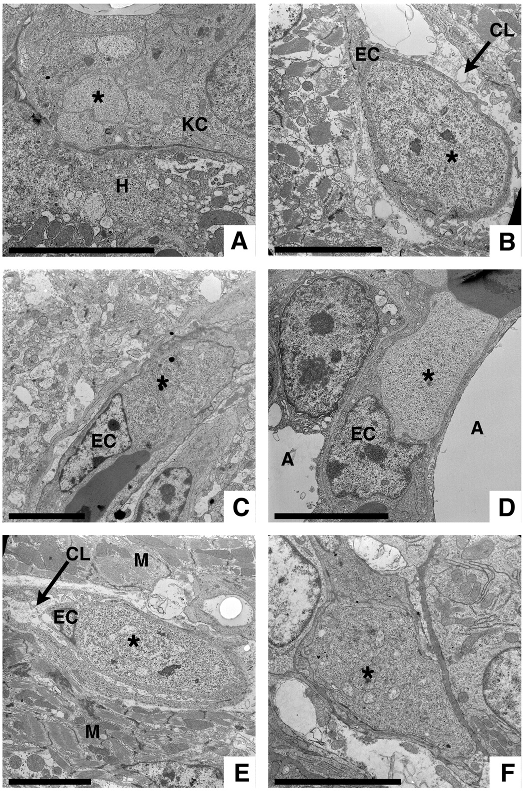
Fig. 3.
Electron microscopic detection of Plasmodium gallinaceum in various chicken organs. Exoerythrocytic stages (*) at different stages of maturation can be found in A) Kupffer cells of the liver, B) endothelial cells (EC) from spleen, C) brain, D) lung, and E) heart as well as in F) an unidentified kidney cell. H = hepatocyte, A = alveolar space, M = myocyte, CL = capillary lumen. Bars = 5 µm.

