Fig. 4.
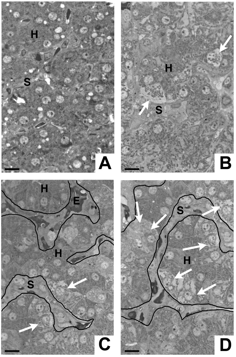
Focal hepatocyte necrosis coincides with cellular occlusion of the sinusoidal lumen. A) Uninfected chick liver exhibiting normal tissue architecture with intact hepatocytes and narrow sinusoids. B–D) Livers from chicks infected by mosquito bite on 8 consecutive days. Note the hydropic swelling and necrotic disintegration of individual hepatocytes (B, arrows) or groups of hepatocytes (C, D; arrows). Patches of necrotic hepatocytes are typically found adjacent to widened sinusoids, which are occluded by infiltrating cells. Note the paucity of erythrocytes in the affected areas. C, D) For clarity, the outline of the sinusoids is visualized by lines. H = hepatocyte, S = sinusoid, E = erythrocyte. Bars = 5 µm.

