
Figure 4. Cell subsets and IL-10.
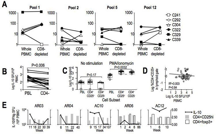
A. HCV-specific (to Pools 1 (Core) and 2 (NS3)), C. albicans-specific, and PHA-induced IL-10 SFU/106 PBMC in unfractionated PBMC and CD8+ T-cell-depleted PBMC in 6 chronic HCV patients. Depleted subset SFU were corrected for changes in T-cell frequency by multiplying by [%CD3PBMC}/[%CD3CD8−]. B. Summed HCV-specific IL-10 SFU/106 PBMC to 12 pools of 15mer overlapping peptides spanning Core, NS3-NS5 with unfractionated and CD4+ T-cell-depleted PBMC in 17 chronic HCV patients. C. IL-10 secretion measured by cytokine bead array (expressed in log IL-10 pg/ml) in 24 hour unstimulated and PMA/ionomycin stimulated cultures of unfractionated PBMC, bead-selected CD4+CD25+ and CD4+CD25− subsets in 5 chronic HCV patients. D. Correlation of frequency of peripheral CD4+CD25−FITC+ T-cells and IL-10 SFU/106 PBMC in 59 chronic HCV patients. E. CD4+CD25hi and CD4+foxp3+ T-cell frequency in acute HCV patients. CD25+ cutoff was defined by 99.9% isotype in the lymphoid gate, while CD25hi was defined by 99.9% of CD8+ T-cells. An example of the gating strategy for CD4+CD25hi population and CD4+foxp3+ gating is shown in Supplementary Figure 1. For 3 AR and 2 AC subjects CD4+CD25hi (grey bars) and CD4+foxp3+ (white bars) are plotted relative to IL-10 SFU/106 PBMC (black line).