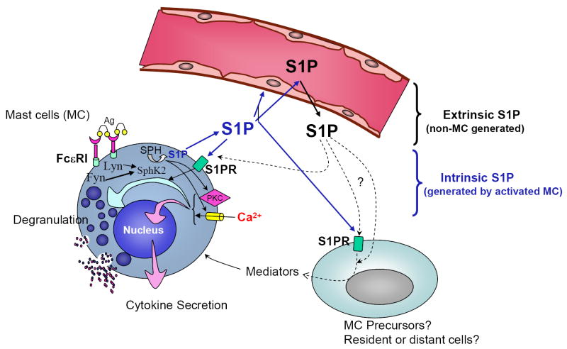
FIGURE 4. In vivo S1P networks affect mast cell function and anaphylaxis.
This figure depicts the mast cell in its physiological environment, and the partnership between extrinsic S1P generated by cells other than mast cells (black arrows; “extracellular compartment” in Figure 2), and intrinsic S1P or S1P generated by stimulated mast cells (blue arrows; corresponding to the “intracellular compartment” in Figure 2), in the regulation of mast cell responsiveness. Loss of SphK2 dramatically reduces the intracellular levels of S1P in mast cells, while loss of SphK1 has no effect. In contrast, deficiency in SphK1 results in reduced levels of S1P in circulation while loss of SphK2 increases the levels. Mast cells may be affected by those changes in different ways. It is possible that changes in S1P in the circulation have a domino effect on the interstitial levels of S1P in tissues, particularly on cells in the proximity of blood vessels where mast cells are present. Changes in those levels may directly affect the priming of S1P2 in the mast cell, impacting on degranulation once the cells are activated. This possibility implies interference of extrinsic S1P with the autocrine loop of S1P2 activation by intrinsic S1P. Other possibilities include an effect of extrinsic S1P on mast cell precursors in the blood stream or mast cells in tissues that will change their phenotypic outcome towards a more or less responsive phenotype. Constant exposure to higher or lower levels of S1P could also alter the phenotype of immune or non-immune cells inducing the generation of mediators that secondarily might influence the differentiation of mast cells. As a consequence of these direct or indirect effects of circulating S1P, mast cells numbers or their phenotypic outcome could be modified.

