
Figure 2.
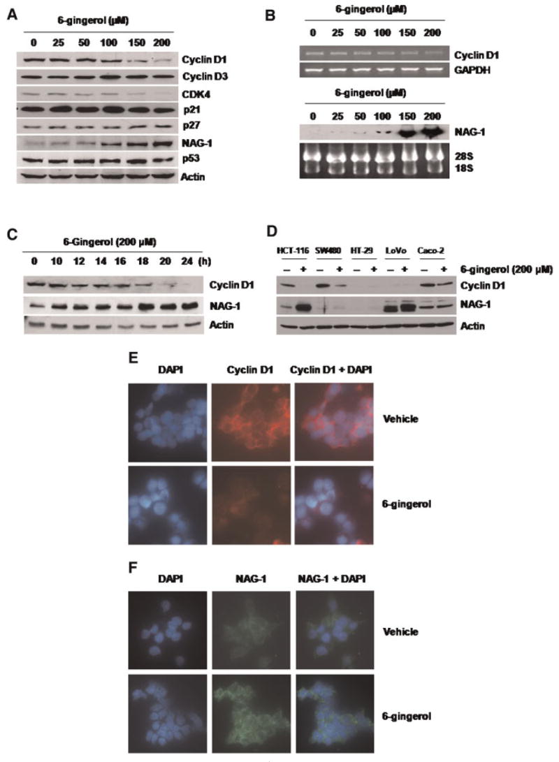
6-Gingerol suppresses cyclin D1 expression and increases NAG-1 expression in HCT-116 cells. (A) HCT-116 cells were treated with the indicated concentration of 6-gingerol for 24 h. Total cell lysates were harvested, and subsequently, 30 μg of total cell lysates were subjected to 14% SDS–PAGE. Cyclin D1, cyclin D3, CDK4, p21, p27, NAG-1, p53, and actin antibodies were probed. The results shown here are representative of three independent experiments. (B) HCT-116 cells were treated with the indicated concentrations of 6-gingerol for 24 h, and then RT-PCR (cyclin D1) or Northern blot analysis (NAG-1) was performed as described in Materials and Methods section. The GAPDH and 28S/18S shown represent a loading control. (C) HCT-116 cells were treated with 200 μM of 6-gingerol at indicated time points. Total cell lysates were harvested, and Western analysis was performed for cyclin D1, NAG- 1, and Actin antibodies. (D) HCT-116, SW480, HT-29, LoVo, and Caco-2 cells were grown as described in Materials and Methods section, and treated with 200 μM of 6-gingerol for 24 h. Western analysis was performed for cyclin D1, NAG-1, and Actin antibodies. (E and F) HCT-116 cells were grown in a glass slide chamber and then treated with 200 μM of 6-gingerol for 24 h. The cells were incubated with a specific antibody for cyclin D1 (1:250) or NAG-1 (1:250) overnight. Cyclin D1 and NAG-1 were detected by using rhodamine conjugate (red) and Alexa Fluor 488 conjugate (green), respectively, and visualized by fluorescence microscopy as described in Materials and Methods section. The DAPI staining (blue) was used to visualize the nuclei of the cells. Magnifications correspond to 400×. [Color figure can be viewed in the online issue, which is available at www.interscience.wiley.com.]