
Figure 4.
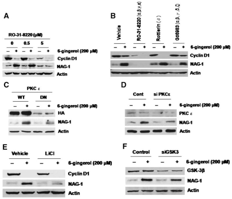
6-Gingerol activates NAG-1 expression via the PKCε and GSK-3β-dependent pathway. (A) HCT-116 cells were pretreated with the indicated concentration of RO-31-8220 for 30 min and then exposed to 200 μM of 6-gingerol for 24 h. Total cell lysates were harvested, and subsequently, 30 μg of total cell lysates were subjected to 14% SDS–PAGE. Cyclin D1, NAG-1, and Actin antibodies were probed. (B) HCT-116 cells were pretreated with 5 μM of RO-31-8220, 0.5 μM of Rottlerin, or 0.5 μM of Gö6983 for 30 min and then exposed to 200 μM of 6-gingerol for 24 h. Western analysis was performed for cyclin D1, NAG-1, and Actin antibodies. (C) HCT-116 cells were transfected with wild type (WT), or dominant negative PKCε expression vector (DN) as described previously [36]. The cells were then treated with 200 μM of 6-gingerol for 24 h. Western analysis was performed for hemagglutinin (HA), NAG-1, and Actin antibodies. (D) HCT-116 cells were transfected with control or PKCε siRNA as described in Materials and Methods section. Then, the cells were treated with 200 μM of 6-gingerol for 24 h. Western analysis was performed using 60 μg of total cell lyates for PKCε and 30 μg of total cell lysates for NAG-1 and Actin antibodies. (E) HCT-116 cells were pretreated with 20 mM of LiCl for 30 min and then exposed to 200 μM of 6-gingerol for 24 h. Western analysis was performed for cyclin D1, NAG-1, and actin antibodies. (F) HCT-116 cells were transfected with control or GSK-3 siRNA. Then, the cells were treated with 200 μM of 6-gingerol for 24 h. Western analysis was performed for GSK-3β, NAG-1, and actin antibodies.