Figure 1.
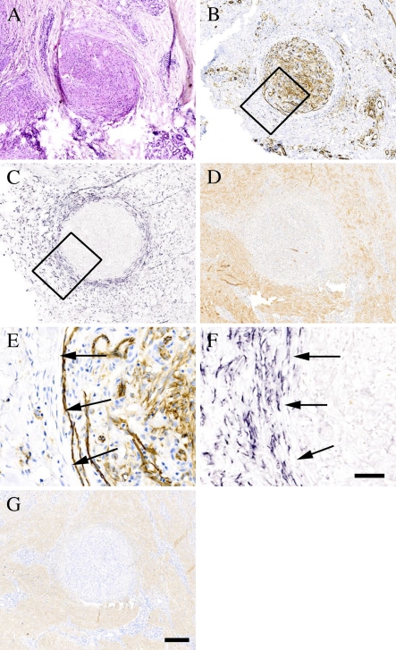
Lack of decorin expression by highly vascularized Kaposi's sarcoma. ICC and ISH analyses of serial sections of a representative Kaposi's sarcoma tissue specimen. (A) Hematoxylin and eosin (HE) staining. (B) Staining with an antibody to the endothelial cell marker CD31. (C) ISH for human decorin. (D) Distribution of decorin identified with LF-136 antiserum for human decorin. Boxed regions shown in B and C represent corresponding areas in serial tissue sections of the same specimen, and magnified illustrations of these regions are shown in E and F, respectively. (G) Distribution of type I collagen identified with LF-67 antiserum for human type I collagen. ICC reactions are brown and, counterstain for nuclei by hematoxylin is blue. Positive digoxigenin (DIG) reaction in ISH assay can be seen in purple. Arrows (E,F) indicate the border between Kaposi's sarcoma and surrounding tissue. Bars: A–D,G = 200 μm; E,F = 50 μm.

