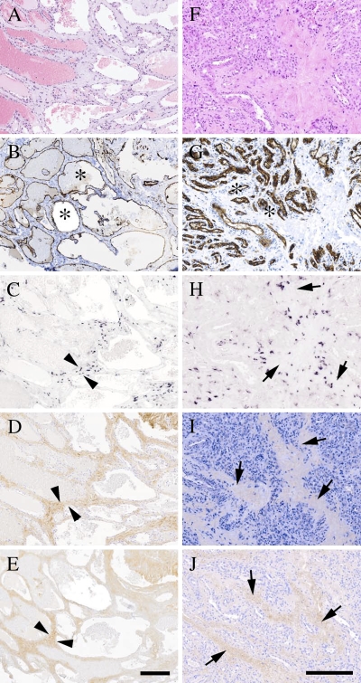
Figure 3.
Cavernous and capillary hemangiomas show positivity for decorin mRNA and also have immunoreactivity for decorin. ICC and ISH analyses of serial sections of representative cutaneous cavernous (A–E) and capillary (F–J) hemangioma tissue specimens. (A,F) HE staining. (B,G) Staining with an antibody to the endothelial cell marker CD31. (C,H) ISH for human decorin. (D,I) Distribution of decorin identified with LF-136 antiserum for human decorin. (E,J) Distribution of type I collagen indentified with LF-67 antiserum for human type I collagen. Both types of hemangiomas contain numerous blood vessel structures (asterisks in B,G). Arrowheads (C–E) indicate border of the connective tissue stroma surrounding selected intratumoral blood vessels in cavernous hemangioma. Arrows (H–J) indicate connective tissue stroma surrounding intratum oral blood vessels in capillary hemangioma. ICC reactions are brown and counterstain for nuclei by hematoxylin is blue. Positive DIG reaction in ISH assay can be seen in purple. Bar (A–E and F–J) = 200 μm.

