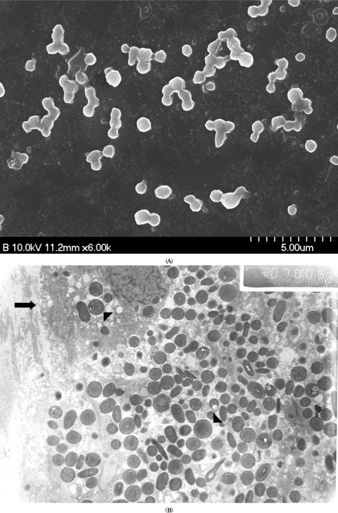
FIGURE 3.
Electron microscopic findings of explanted intraocular lens (IOL) and residual capsule in two patients with coinfection of O. anthropi and P. acnes. (A) Scanning electron microscopy of a patient (No. 9) reveals polymorphic and rod-shaped bacillus at the surface of explanted IOL. (B) Transmission electron microscopy of a patient (No. 8) also shows the polymorphic organisms with diverse sizes (arrowhead) and a pathogen-laden macrophage (arrow).

